

BIOS 242 Week 5 Immune and Lymphatic system Lab
Student Name
Chamberlain University
BIOS-242 Fundamentals of Microbiology
Prof. Name
Date
Lab Overview
This laboratory exercise is divided into two key components. The first part focuses on identifying and labeling the anatomical structures of the lymphatic system. This section helps learners visually connect the structures with their physiological roles. The second part involves answering a set of reflective questions to reinforce theoretical understanding of the immune and lymphatic systems. Once all sections are completed, students must submit their responses to the designated submission area for evaluation.
Part 1: Anatomical Structures of the Lymphatic System
The following table outlines the primary anatomical structures of the lymphatic system along with their assigned labels:
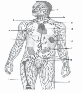
| Anatomical Structure | Label |
|---|---|
| Tonsils | a |
| Cervical Lymph Nodes | b |
| Thoracic Duct | c |
| Thymus | d |
| Spleen | e |
| Peyer’s Patches (in Intestine) | f |
| Inguinal Lymph Nodes | g |
| Lymphatic Vessels | h |
| Bone Marrow | i |
| Cisterna Chyli | j |
| Axillary Lymph Nodes | k |
| Right Lymphatic Duct | l |
Part 2: Questions and Answers
Explain why the lymphatic system is a one-way system, whereas the blood vascular system is a two-way system.
The lymphatic system is a unidirectional network designed to collect excess interstitial fluid and filter it through lymph nodes before returning it to the circulatory system. This ensures that waste products, pathogens, and cellular debris are processed before re-entering the bloodstream. In contrast, the blood vascular system is bidirectional; arteries deliver oxygen and nutrients to tissues, while veins return deoxygenated blood and waste products back to the heart for reoxygenation and filtration.
How do lymphatic vessels resemble veins?
Lymphatic vessels share several similarities with veins. Both have thin walls and rely on external forces such as skeletal muscle contractions and pressure changes from respiration to move fluid. Like veins, lymphatic vessels contain valves that prevent backflow, ensuring the lymph flows only toward the thoracic region. However, lymphatic vessels tend to have thinner walls and lower pressure compared to veins.
How do lymphatic capillaries differ from blood capillaries?
Lymphatic capillaries differ from blood capillaries in both structure and function. Blood capillaries connect arterioles and venules, allowing for the exchange of gases, nutrients, and waste products. Lymphatic capillaries, on the other hand, collect interstitial fluid, proteins, lipids, and immune cells, transporting them into lymphatic venules. Structurally, lymphatic capillaries have larger diameters and more permeable walls, which allow them to absorb larger molecules such as lipids.
BIOS 242 Week 5: Immune and Lymphatic System Lab
What is the function of the lymphatic vessels?
Lymphatic vessels are responsible for transporting lymph from tissues to the venous circulation. They facilitate the removal of cellular waste and pathogens through phagocytosis within lymph nodes, contribute to immune surveillance by transporting lymphocytes, and also absorb dietary fats through specialized vessels known as lacteals in the small intestine.
What is lymph?
Lymph is a clear to slightly yellowish fluid that circulates through the lymphatic system. It contains water, proteins, lipids, immune cells (primarily lymphocytes), and absorbed fats (chyle). Its function is to maintain fluid balance, remove waste, and transport immune cells to areas of infection or inflammation.
What factors are involved in the flow of lymphatic fluid?
The movement of lymph is primarily driven by skeletal muscle contractions (the “milking effect”) and respiratory pressure changes during inhalation and exhalation. Additionally, valves within lymphatic vessels prevent backflow, ensuring unidirectional movement toward the thoracic cavity.
What name is given to the terminal duct draining most of the body?
The Thoracic Duct is the terminal lymphatic vessel that drains lymph from the majority of the body, including both lower limbs, the abdomen, the left thorax, left upper limb, and left side of the head and neck.
What is the function of B cells in the immune response?
B cells are a critical part of humoral immunity. Upon encountering an antigen, B cells differentiate into plasma cells, which produce and secrete antibodies. These antibodies specifically target pathogens, neutralize toxins, and flag invaders for destruction by other immune cells. Memory B cells are also formed, providing long-term immunity against previously encountered pathogens.
What is the role of T cells?
T cells contribute to cell-mediated immunity. Cytotoxic T cells directly destroy virus-infected or cancerous cells, while helper T cells activate B cells and enhance the activity of cytotoxic T cells. Regulatory T cells modulate immune responses to prevent overactivation, thereby maintaining immune system balance.
Define the following term related to the operation of the immune system: Recognition of self from non-self.
Recognition of self from non-self is the immune system’s ability to differentiate between the body’s own cells and foreign invaders. Self-recognition prevents autoimmunity by ensuring the immune system does not attack healthy tissue. Non-self refers to foreign antigens such as bacteria, viruses, or allergens. When previously encountered, memory cells enhance the immune system’s ability to mount a faster and stronger response against these pathogens.
References
Guyton, A. C., & Hall, J. E. (2021). Textbook of medical physiology (14th ed.). Elsevier.
Marieb, E. N., & Hoehn, K. (2019). Human anatomy & physiology (11th ed.). Pearson.
BIOS 242 Week 5 Immune and Lymphatic system Lab
Turgeon, M. L. (2020). Immunology & serology in laboratory medicine (6th ed.). Elsevier.